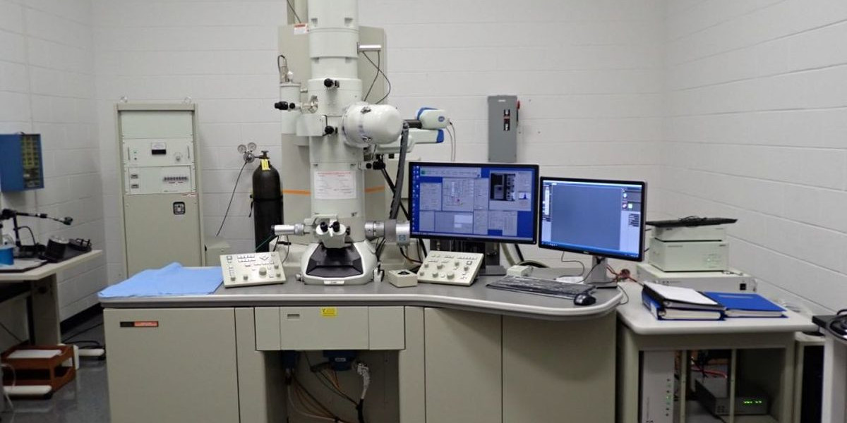
EM microscopy, or electron microscopy, refers to advanced imaging techniques that use beams of electrons instead of light to magnify and resolve tiny details of specimens. Unlike traditional optical microscopes, electron microscopes can achieve resolutions at the nanometer or even atomic scale, making them essential for fields like materials science, nanotechnology, biology, and medicine.
There are two main types of EM microscopy:
Transmission Electron Microscopy (TEM): Electrons pass through an ultra-thin sample to reveal internal structures in high detail.
Scanning Electron Microscopy (SEM): Electrons scan the surface of a specimen, producing highly detailed, 3D-like images of texture and topography.
While EM microscopy offers incredible resolution, it requires a controlled laboratory environment, sample preparation, and expertise. In contrast, portable optical microscopes like the EM1 are designed for fieldwork, providing practical, real-time imaging with magnification up to 1000x.
Both approaches serve different but complementary purposes—EM microscopy for ultra-high-resolution lab analysis, and portable microscopes for accessible, on-site investigations.